


A guide to Sludgy Gallbladder
What is Sludgy Gallbladder?
Welcome to our article on Sludgy gallbladder, in this article we will explain everything you need to know about sludgy gallbladder or to give it, its proper name Gallbladder mucocele. This is a serious condition that affects dogs, characterized by the accumulation of excessive mucus within the gallbladder. It can lead to various complications, including biliary duct obstruction, cholecystitis (inflammation of the gallbladder), and even gallbladder rupture. Early diagnosis and appropriate treatment are crucial for the successful management of this condition. In this comprehensive guide, we will explore the causes, symptoms, diagnosis, and treatment options for canine gallbladder mucocele.
Canine Gallbladder Mucocele
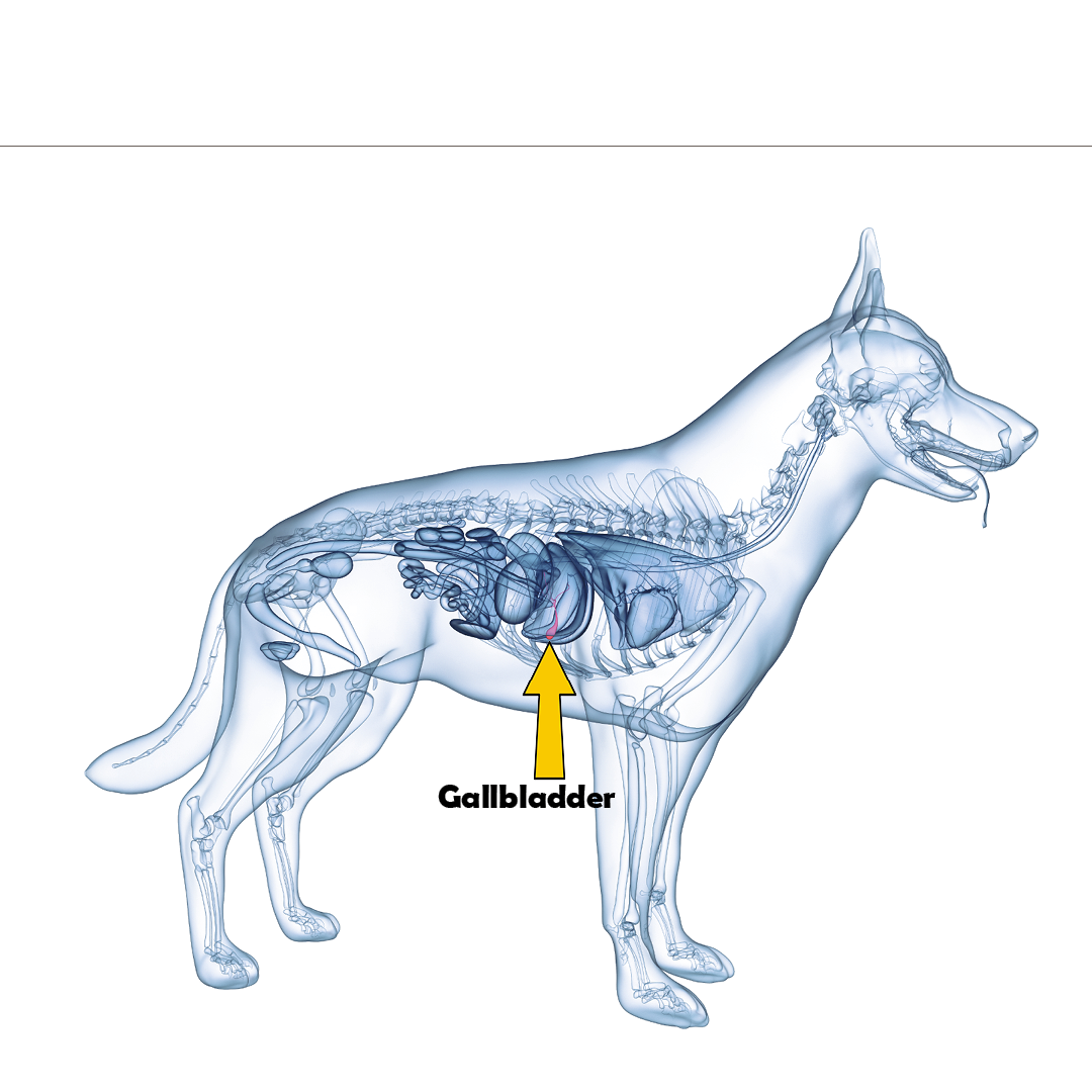
The gallbladder is an essential organ involved in the digestive process of dogs. It stores and fortifies bile, which aids in digestion and the absorption of vital nutrients. Canine gallbladder mucocele occurs when there is an abnormal accumulation of thick, gelatinous bile within the gallbladder. This condition is caused by cystic mucosal hyperplasia and hypersecretion of mucus. Over time, the increased viscosity of the bile leads to the filling of the entire gallbladder, and in some cases, the hepatic and common bile ducts with mucus.
The underlying cause of gallbladder mucocele is still unknown, but genetic predisposition may play a role, with certain breeds like Shetland Sheepdogs, Miniature Schnauzers, Cocker Spaniels, and Border Terriers being more susceptible. Gallbladder mucoceles usually occur in older dogs, and there is no sex predilection. Additionally, this condition has been associated with endocrine diseases such as hyperadrenocortism and hypothyroidism. Dogs with Cushing’s disease are 29 times more likely to develop gallbladder mucocele. Other factors that may contribute to its development include dyslipidemia (imbalance of lipids), the excessive use of corticosteroids, and gallbladder dysmotility (poor motility).
Symptoms of Canine Gallbladder Mucocele
Unfortunately, the clinical signs of gallbladder mucocele in dogs can be vague and non-specific. Some common symptoms include:
- Decreased appetite
- Lethargy
- Vomiting
- Abdominal pain
- Jaundice (yellowing of the skin and eyes).
It’s important to note that not all affected dogs will exhibit all of these symptoms, and the severity of the symptoms can vary. Some dogs may be asymptomatic, while others may experience more severe signs. If you notice any of these symptoms in your dog, it is essential to seek veterinary attention for a proper diagnosis and treatment.
Causes of Canine Gallbladder Mucocele
The exact cause of gallbladder mucocele in dogs is still unknown. However, several factors may contribute to its development. These include gallbladder disease, glucocorticoid therapy, progestational therapy, gallbladder stones, inflammation of the gallbladder, biliary sludge, cystic mucinous hyperplasia, mucus-secreting cell dysfunction, and dysmotility of the gallbladder. Certain conditions and predisposing factors can increase the likelihood of gallbladder mucocele in dogs. These include being a small to medium-sized dog, middle to older age, specific breeds such as Shetland Sheepdogs and Cocker Spaniels, endocrine diseases like Cushing’s disease, high cholesterol, a high-fat diet, pancreatitis, gallbladder disorders, inflammatory bowel disease, and gallbladder cyst formation.
Diagnosis of Canine Gallbladder Mucocele
To diagnose gallbladder mucocele in dogs, a comprehensive approach is necessary. The diagnostic process typically involves a combination of clinical signs, physical examination findings, laboratory analysis, and abdominal imaging. During the physical examination, your veterinarian may observe cranial abdominal pain and icterus. Blood tests, including a complete blood count and biochemistry panel, may reveal elevated liver enzymes (ALT and ALKP), total bilirubin, and leucocytosis. However, these blood markers can be within the reference range, especially in early cases. Abdominal ultrasound is the preferred imaging modality for diagnosing gallbladder mucocele.
The characteristic ultrasonographic finding is a stellate or finely striated pattern within the gallbladder, often resembling a kiwi fruit. It’s important to differentiate gallbladder mucocele from biliary sludge, which can be mistaken for a mucocele. Biliary sludge will move with gravity, while a mucocele will not.
In some cases, gallbladder ruptures can occur before the diagnosis is made. If there is evidence of gallbladder rupture or biliary peritonitis, it is considered an emergency, and immediate surgical intervention is required. An abdominocentesis may be performed to confirm the presence of bile peritonitis. If the common bile duct is suspected to be obstructed, further evaluation and catheterization may be necessary. Bacterial culture and sensitivity testing of the gallbladder wall, as well as a hepatic biopsy, may be recommended to guide treatment and ensure appropriate management.
Treatment of Canine Gallbladder Mucocele
The treatment of canine gallbladder mucocele typically involves surgical removal of the gallbladder, known as cholecystectomy. While medical management with certain medications has been reported, there is limited literature supporting its effectiveness. Therefore, surgical intervention is the preferred and most reliable treatment option. Before surgery, a coagulation profile should be performed to ensure the dog’s blood clotting ability. If coagulation tests indicate abnormalities, administration of vitamin K1 or fresh frozen plasma may be necessary to correct the coagulation status. Broad-spectrum intravenous antibiotics should also be administered peri-operatively to prevent infection. Laparoscopic cholecystectomy may be performed in selected cases where there is no evidence of gallbladder rupture or extrahepatic biliary obstruction. However, open celiotomy is the more common approach for cholecystectomy in most cases.
The prognosis following cholecystectomy for gallbladder mucocele is generally favourable. Most dogs will make a full recovery and return to normal activity. However, complications can occur, including biliary peritonitis, pancreatitis, and re-obstruction of the common bile duct with congealed mucus or bile. The peri-operative mortality rate is reported to be around 20%, but with appropriate management and care, this rate can be significantly lower. Long-term recurrence is rare in dogs that survive the peri-operative period.
Incidental Gallbladder Mucocele: What to Do?
The management of incidental gallbladder mucoceles can be challenging. Incidental gallbladder mucoceles refers to cases where the mucocele is discovered incidentally during diagnostic tests or procedures performed for other reasons. It is important to differentiate between early mucoceles and gallbladder sludge, as sludge is a common incidental finding and does not require immediate intervention.
However, if a gallbladder mucocele is confirmed, surgical intervention is usually recommended unless there are specific contraindications. Delaying surgical management in these cases can increase the risk of progression, gallbladder rupture, and secondary complications. Therefore, it is crucial to consult with a veterinarian and discuss the appropriate course of action based on the individual case.
Canine gallbladder mucocele is a serious condition that requires prompt diagnosis and treatment. The accumulation of excessive mucus within the gallbladder can lead to various complications and even be life-threatening if left untreated. If you notice any symptoms or suspect gallbladder mucocele in your dog, it is important to seek veterinary attention immediately.
We hope you have found our article on Sludgy gallbladder interesting and useful. Here is an article by the American College of Veterinary Surgeons on Gall bladder mucocele, for further reading.
Disclaimer: The contents of Itsadogz.life website are for informational and educational purposes only and should NOT replace professional Veterinary Advice. Always consult a qualified Veterinarian regarding your dog’s health and any concerns you may have.
Neither the owners nor creators of this site are engaged in providing veterinary medical services via this site and assume no responsibility for any consequence relating directly or indirectly to any action you take or do not take based on the information, services, products, or other materials provided on this site.
