


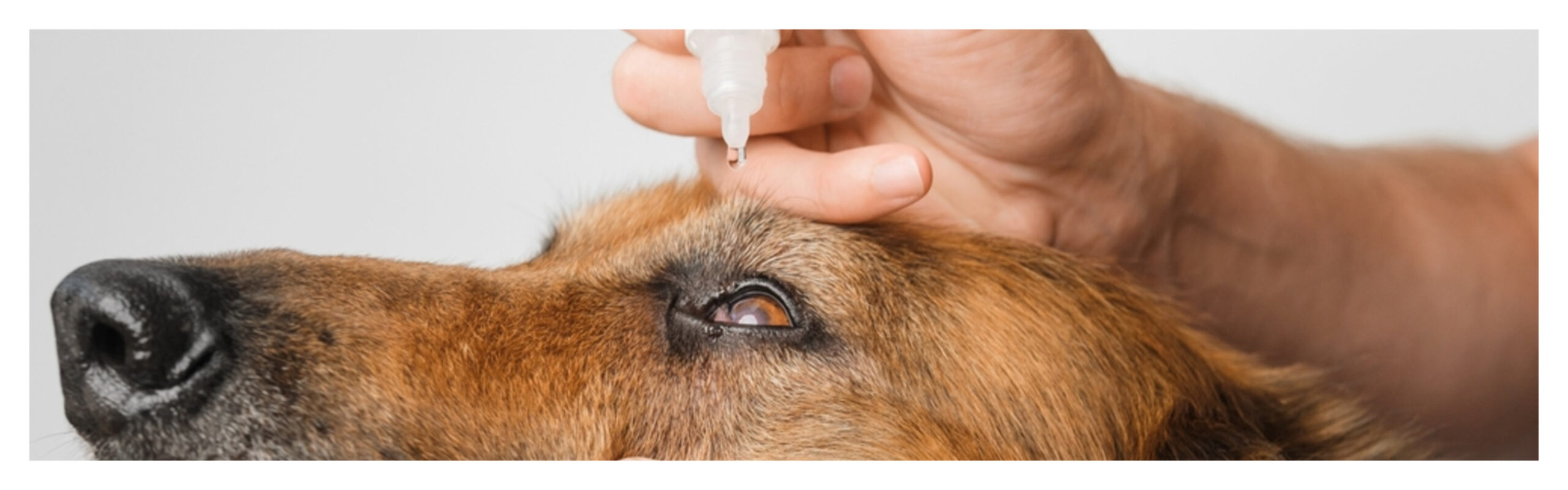
A Guide From Diagnosis to Recovery
Horner’s syndrome in dogs (also called oculosympathetic paresis) is a neurological condition that affects the eye and facial muscles on one side of your dog’s face. The condition most commonly appears in dogs between five and eight years old, with certain breeds like Labrador Retrievers, Golden Retrievers, Dobermans, Weimaraners, Shetland Sheepdogs and Collies showing a higher incidence than others.
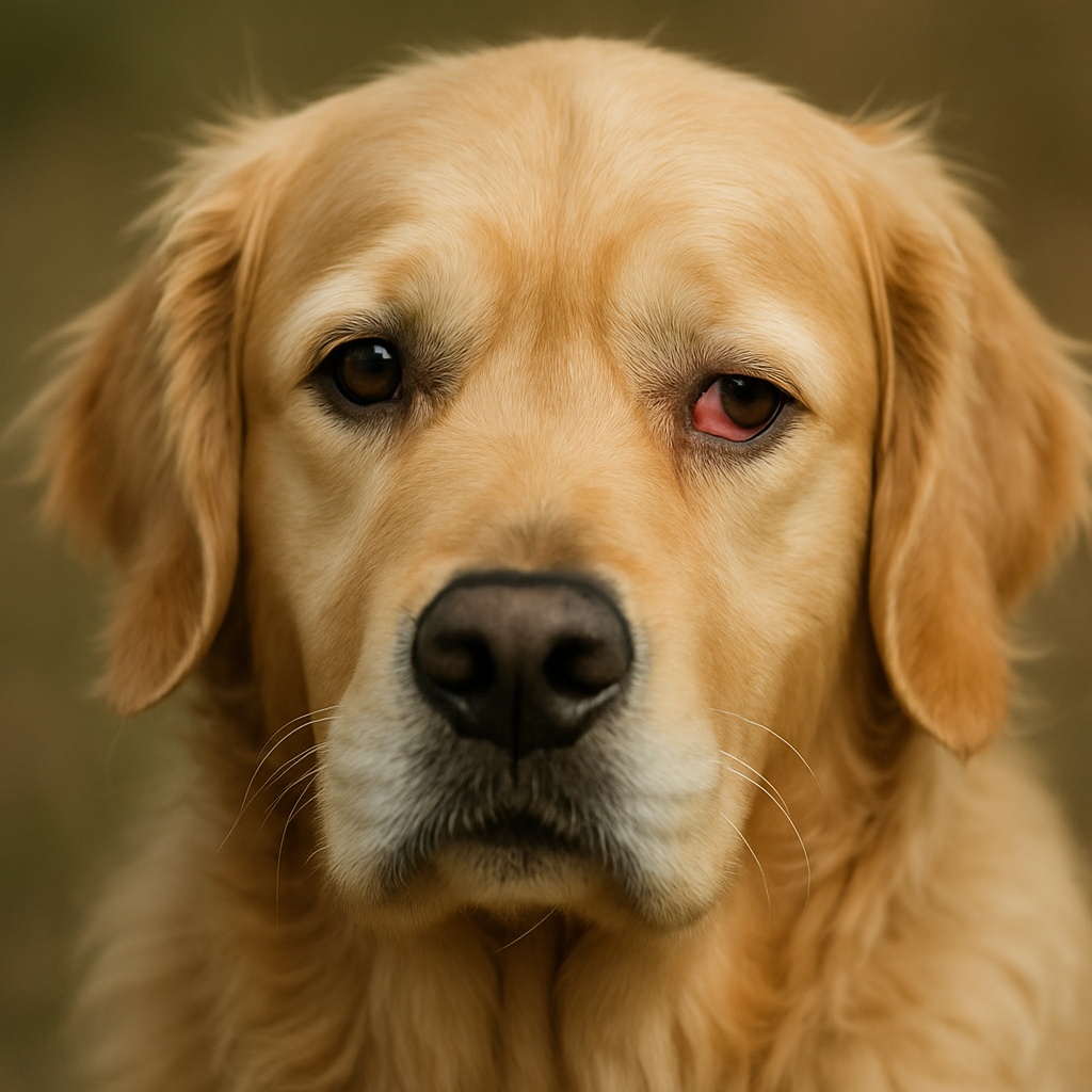
The syndrome occurs when there’s damage to the sympathetic nerves that control specific eye muscles. When these nerves aren’t working properly, you’ll notice several distinctive changes to your dog’s appearance:
- Drooping of the upper eyelid (ptosis)
- A constricted pupil (miosis)
- The eye appears sunken (enophthalmos)
- The third eyelid becomes unusually visible
This last symptom – the visible third eyelid – often causes the most concern for dog owners, as it creates a prominent whitish-pink membrane across part of the eye.
Horner’s syndrome can be tricky to diagnose because in approximately half of all cases, vets cannot determine what caused the nerve damage. The most common identifiable causes include neck injuries, ear infections, and occasionally more serious conditions like tumours affecting the nervous system. Most cases do resolve on their own, though recovery typically takes anywhere from 16 weeks to 6 months, depending on the underlying cause and severity.
This guide will help you understand how Horner’s syndrome is diagnosed, the available treatment options, and what to expect during your dog’s recovery.
How Horner’s Syndrome Affects Your Dog’s Eye and Face
Horner’s syndrome happens when the sympathetic nerve pathway controlling your dog’s eye muscles gets damaged or interrupted. This creates the distinctive one-sided facial changes you’ll notice.
Understanding the Role of the Sympathetic Nervous System
The sympathetic nervous system controls many automatic body functions, including how your dog’s pupils dilate, where the eyelids sit, and how the eye positions itself in the socket. This system operates through a three-neuron pathway that originates in the brain, travels down the spinal cord, and terminates at the eye muscles. When any part of this pathway gets damaged, the nerve signals can’t reach the eye properly, which causes the characteristic signs of Horner’s syndrome.
Think of it like a telephone line – if there’s a break anywhere along the wire, the message doesn’t get through to the other end.
The Main Symptoms: Miosis, Ptosis, and Enophthalmos
Horner’s syndrome typically affects only one side of your dog’s face, creating an obvious asymmetry that can be quite striking. The three main symptoms each have specific medical names:
Miosis is the constricted pupil you’ll see compared to the normal eye. This happens because the muscles that usually make the pupil larger aren’t getting the nerve signals they need to work properly.
Ptosis refers to the drooping upper eyelid that gives your dog a sleepy appearance on the affected side. The muscle that normally holds the eyelid open loses its nerve supply, so it relaxes and drops.
Enophthalmos is the sunken appearance of the eye within its socket. The muscles that usually keep the eye positioned properly in the skull also relax when they lose their nerve signals, causing the eye to sink back slightly.
These symptoms can appear quite suddenly and may be alarming because they create such a noticeable change in your dog’s facial appearance.
Why the Third Eyelid Becomes Visible
The prominent third eyelid (also called the nictitating membrane) is often what worries owners most. Normally, this translucent structure stays tucked away at the inner corner of your dog’s eye, only appearing partially when they’re asleep or when the eye needs protection from something irritating.
When Horner’s syndrome develops, the muscles that usually keep this membrane retracted stop working properly. The third eyelid then slides across part of the eye, creating what looks like a whitish-pink film covering the lower portion of the eyeball. Combined with the other facial changes, this gives the eye a distinctly abnormal appearance that’s quite characteristic of the condition.
What causes Horner’s syndrome?
Determining what’s causing your dog’s Horner’s syndrome can be challenging. The condition stems from damage to the sympathetic nerves that control the eye muscles, but the underlying triggers vary significantly between cases.
Ear infections – the most common identifiable cause
Ear infections represent the most common identifiable cause of Horner’s syndrome in dogs. Middle or inner ear disease (otitis media or interna) damages the nerve fibres close to the ear, creating what vets call “third order lesions.” These cases often appear alongside other symptoms you might recognise – head tilting, loss of balance, or your dog seeming dizzy or disoriented (vestibular disease).
Trauma and injuries
Trauma to the neck or chest area can damage the sympathetic nerve pathway as it travels from the spinal cord to the eye. This might happen from bite wounds, blunt force trauma, or even pulling too hard on a collar lead. Foreleg injuries can also cause problems, particularly if the limb gets pulled and stretches the nerves where they exit the spinal cord in the armpit region. These injuries typically affect what specialists call the “preganglionic segment” of nerves.
Serious neurological conditions
More concerning causes include problems affecting the brain, brainstem, or spinal cord itself. These “first order lesions” can result from brain tumours, strokes, spinal strokes (fibrocartilaginous emboli), or intervertebral disc disease in the neck region. with Horner’s syndrome. When vets suspect these types of causes, advanced imaging like MRI scans becomes necessary to get a proper diagnosis. Cervical myelopathy has been observed in approximately 10% of dogs
Idiopathic cases – when no cause can be found
Here’s where it gets interesting – in approximately half of all cases, vets cannot identify any underlying cause despite thorough examination. These are called “idiopathic” cases. Golden Retrievers seem particularly prone to this type, with an incidence of 2.6% versus 0.03% in other breeds. Collies may also face increased risk. The good news is that idiopathic cases generally carry a better prognosis, with many resolving on their own within several weeks.
The mystery surrounding these idiopathic cases highlights why Horner’s syndrome can be so frustrating to diagnose, but it also means that many dogs recover without needing extensive treatment.
How is Horner’s Syndrome Diagnosed?
Diagnosing Horner’s syndrome requires your vet to work through a systematic process to confirm the condition and identify what’s causing it. When you notice the distinctive eye changes in your dog, a veterinary consultation becomes essential for proper evaluation.
What Happens During the Initial Examination
Your vet will start with a thorough physical examination, paying particular attention to both eyes under different lighting conditions. This helps determine which pupil is actually abnormal, as the less mobile pupil typically indicates the problem. They’ll also need to rule out other potential causes of pupil constriction (miosis), including conditions like uveitis, endophthalmitis, panophthalmitis, and ulcerative keratitis.
The examination often includes a Schirmer Tear Test to measure tear production and a fluorescein stain to check for any corneal damage. Don’t be surprised if your vet spends considerable time examining your dog’s ears and neck area, as these are common sources of the nerve damage that causes Horner’s syndrome.
Phenylephrine Eye Drops Test
Your vet might use a phenylephrine response test to pinpoint where along the nerve pathway the damage has occurred. While remains the gold standard for confirming Horner’s syndrome diagnosis, phenylephrine testing is more practical and widely available cocaine solution (5-10%).
When 1% phenylephrine drops are applied to the affected eye, the timing of the response tells your vet important information about the location of nerve damage. If the pupil dilates within 20 minutes, this suggests postganglionic nerve damage (often from ear infections). A minimal or delayed response indicates preganglionic or central nerve damage. This distinction helps narrow down potential causes significantly.
Additional Tests Your Vet May Recommend
Depending on the initial findings, your vet might suggest several diagnostic tests to identify underlying conditions. These typically include baseline blood work, a thorough ear examination with an otoscope, and that include the neck region.
For cases where central or preganglionic nerve damage is suspected (particularly if your dog shows other neurological symptoms), advanced imaging such as MRI or CT scans becomes necessary to evaluate the entire nerve pathway through the brain, spinal cord, mediastinum, neck, middle ear, and orbit.
When Specialist Referral Becomes Necessary
Your vet might recommend seeing a veterinary neurologist if they suspect first-order lesions affecting the brain, brainstem, or spinal cord. This is particularly likely if your dog shows additional neurological symptoms beyond the eye changes, or if initial tests reveal potential tumours, vascular accidents, or spinal cord issues.
Veterinary neurologists have specialised equipment and expertise to perform advanced procedures including cerebrospinal fluid analysis or biopsies of suspicious masses. Don’t worry if a referral is suggested – it simply means your vet wants to ensure your dog receives the most appropriate care for their specific situation.
Treatment, Recovery, and Home Care
Treatment for Horner’s syndrome focuses on addressing the underlying cause rather than the syndrome itself, which is fortunately not painful and doesn’t interfere with your dog’s vision.
How is Horner’s Syndrome treated?
Your vet’s treatment approach will depend entirely on what’s causing the condition. If ear infections are to blame, antibiotics and anti-inflammatory medications will likely be prescribed. When trauma is responsible, treatment centres on reducing inflammation and giving damaged nerves time to heal properly. More serious causes like tumours might require surgical intervention.
What’s particularly interesting is that and many of these resolve spontaneously within several weeks (this is actually good news, as it means the prognosis is often quite favourable), about half of all cases have no identifiable cause
Eye Drops and Supportive Care
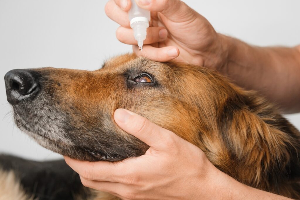
Phenylephrine eye drops are commonly prescribed to temporarily improve the appearance of symptoms. These drops help reduce the prominence of the third eyelid and assist with pupil dilation, providing both cosmetic improvement and better vision whilst the underlying issue resolves. Your vet might also recommend artificial tears if your dog shows decreased tear production (this helps keep the eye comfortable during recovery).
What to expect during recovery
Recovery times vary considerably depending on the underlying cause. For idiopathic cases (where no cause can be found), improvement typically begins within several weeks, with . You’ll notice gradual changes during this time – the third eyelid usually retracts first, followed by improvements in the drooping eyelid and the sunken appearance. A complete resolution occurring within 16 weeks to 6 months.
Prevention and ongoing management
Regular ear cleaning and checks can help prevent the ear infections that sometimes trigger Horner’s syndrome. Using harnesses instead of collar leads also helps prevent neck injuries that might damage the sympathetic nerves. Most importantly, follow your vet’s recommendations for managing any identified underlying conditions to reduce the risk of recurrence.
Horner’s syndrome can look alarming when you first notice the changes to your dog’s face, but the condition itself doesn’t cause pain or affect your dog’s vision. Most dogs cope very well with the symptoms whilst their nerves heal.
The good news is that many cases resolve on their own over time. Your vet will help determine if there’s an underlying cause that needs specific treatment, such as ear infections or injuries that require attention. Recovery takes patience – you’ll typically see the third eyelid return to normal first, followed by gradual improvement in the drooping eyelid and sunken appearance.
Don’t be afraid to ask your vet questions about your dog’s recovery progress or if you notice any changes during the healing process. Regular check-ups help ensure any underlying conditions are properly managed and allow your vet to monitor improvement.
Simple prevention measures can reduce the risk of Horner’s syndrome recurring. Keep your dog’s ears clean and check them regularly for signs of infection. Use a harness rather than a collar when walking your dog, as this reduces the risk of neck injuries from pulling or sudden movements.
Most dogs with Horner’s syndrome go on to make a full recovery, though it can take several months. The condition generally has a good outlook, particularly for cases where no underlying cause is found. With proper veterinary care and monitoring, your dog can return to their normal, happy self.
Key Takeaways
Understanding Horner’s syndrome helps you recognise when your dog needs veterinary attention and what to expect during treatment.
- Horner’s syndrome causes distinctive one-sided facial symptoms: drooping eyelid, constricted pupil, sunken eye, and visible third eyelid
- Approximately half of cases have no identifiable cause and resolve naturally within 16 weeks to 6 months
- Ear infections are the most common identifiable trigger, making regular ear care essential for prevention
- The condition is typically painless and doesn’t affect vision, though prompt veterinary diagnosis remains crucial
- Treatment focuses on addressing underlying causes rather than the syndrome itself, with supportive eye drops providing comfort
Most dogs with Horner’s syndrome experience gradual improvement as damaged nerves heal, making the prognosis generally favourable with proper veterinary care.
Disclaimer: The contents of Itsadogz.life website are for informational and educational purposes only and should NOT replace professional Veterinary Advice. Always consult a qualified Veterinarian regarding your dog’s health and any concerns you may have.
Neither the owners nor creators of this site are engaged in providing veterinary medical services via this site and assume no responsibility for any consequence relating directly or indirectly to any action you take or do not take based on the information, services, products, or other materials provided on this site.
